Hypercalcemia
Master the patient approach to hypercalcemia through our interactive clinical case with clear visual guides.
CALCIUM AND BONE
Case Presentation
A 58-year-old man is referred to your outpatient clinic by his primary care physician for evaluation of elevated serum calcium. The referral notes report a slight fatigue over the past six months, which prompted a thorough medical workup, including the calcium measurement. His past medical history includes hypertension and a conservatively managed episode of urolithiasis three years ago. He is currently taking Lisinopril 10 mg daily for blood pressure control and no other medications.
Laboratory test 2 months ago
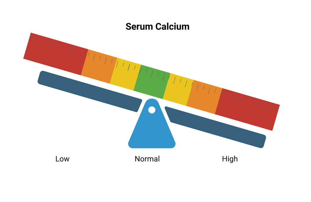
Serum calcium level: 11.8 mg/dL (Reference Range: 8.5–10.2 mg/dL) or 2.94 mmol/L (Reference Range: 2.20 - 2.55 mmol/L)
Serum albumin: 41 g/L (Reference Range: 34-50 g/L)
Serum creatinine: 75 µmol/l (Reference Range 45 - 84 µmol/L)
eGFR: > 90 ml/min/1.73 m² (Reference Range >59 ml/min/1.73 m²)
Laboratory test 1 month ago
Serum calcium level: 11.9 mg/dL (Reference Range: 8.5–10.2 mg/dL) or 2.97 mmol/L (Reference Range: 2.20 - 2.55 mmol/L)
Referral & Emergency Evaluation
To make optimal use of the patient’s history, we will first review the possible etiologies of this patient’s hypercalcemia.
History
The patient denies any history of fractures or bone pain. Urolithiasis was managed conservatively 3 years ago without any subsequent episodes. The stone was not examined. He denies any respiratory symptoms like dyspnea and cough, as well as B-Symptoms like weight loss or nighttime sweating. Regarding his medication history, he confirms that he is not taking any diuretics, lithium, or excessive amounts of calcium or vitamin D supplements. Additionally, there is no known family history of endocrine disorders, jaw tumours or disorders associated with multiple endocrine neoplasia.
Clinical Exam
On examination, his vital signs are within normal limits, with a blood pressure of 124/78 mmHg and a heart rate of 76 bpm. He appears well-hydrated, with no immediate signs of distress. No lymphadenopathy is noted. Cardiopulmonary examination is uneventful as well as clinical examination of the neck and skin.
Diagnosis
Results of the patient
Ionized Calcium: 6.2 mg/dl or 1.55 mmol/L (Reference Range: 4.4-5.2 mg/dL or 1.10-1.30 mmol/L)
PTH: 105 pg/ml (Reference Range: 15–65 pg/mL)
Phosphate: 0.82 mmol/L (Reference Range: 0.81 - 1.50 mmol/L)
25OHD: normal
Serum creatinine: 75 µmol/l
eGFR: > 90 ml/min/1.73 m²
Management
The neck ultrasound reveals a hypoechoic, well-circumscribed lesion measuring approximately 1.3 x 0.8 cm, located posterior to the inferior pole of the right thyroid lobe. The lesion appears distinct from the thyroid gland, with increased vascularity predominantly at the periphery and polar regions on Doppler imaging, suggestive of a parathyroid adenoma. No significant cervical lymphadenopathy is observed, and the thyroid gland itself appears normal in size and echotexture.
Follow-up imaging with 18F-fluorocholine PET/CT demonstrates a metabolically active nodule at the corresponding site. No additional hypermetabolic foci are identified in the remaining parathyroid glands, and there is no evidence of ectopic parathyroid tissue.
The patient agrees with a referral to a parathyroid surgeon. The following are knowledge checkpoints, questions, and exercises designed to strengthen understanding and clinical management of patients with primary hyperparathyroidism. After that we will revisit our patient’s case.
The patient’s bone mass density (BMD) shows T-scores of -0.8 at the lumbar spine, -1.0 at the femoral neck, and -1.6 at the distal one-third of the radius.
Renal function is normal. The 24-hour urinary calcium excretion is 280 mg/day
Kidney ultrasound revealed a 2 mm stone in the upper segment of the renal calyx, without signs of ureteral stones, kidney congestion, or hydronephrosis.
You conclude, that this patient does not meet the criteria for targeted genetic testing of hereditary primary hyperparathyroidism.
The patient decides to undergo parathyroidectomy. A parathyroidectomy of the right inferior parathyroid gland is planned.
The suspected adenoma located posterior to the right inferior thyroid lobe was identified during surgery. The parathyroid gland posterior to the right superior thyroid lobe was also visualized, showing no signs of adenomatous tissue. Intraoperative PTH levels dropped to 22 pg/ml, indicating successful removal of the hyperfunctioning gland with no evidence of additional overactive tissue.
PTH is measured at 11 pg/ml with total Calcium levels at 8.4 mg/dl (2.1 mmol/L) three hours after surgery with without any symptoms of hypocalcemia.
Clinical examination of the surgical site revealed no evidence of postoperative hematoma. The patient reported no changes in voice quality, and fiberoptic laryngoscopy confirmed intact laryngeal nerve function without signs of nerve injury.
Histopathological analysis confirmed a well-circumscribed, encapsulated lesion composed of uniform chief cells arranged in nests and trabeculae, with reduced adipose tissue and no capsular or vascular invasion, consistent with a parathyroid adenoma.
The patient was started on treatment with 1 g of calcium carbonate twice daily and calcitriol 0.25 mcg twice daily. Subsequent calcium levels remained stable, and the patient was discharged.
Seven days after surgery, the patient reports no symptoms of hypocalcemia. PTH is 13 pg/ml and calcium levels are within the normal range. You decide to continue the current treatment and schedule a follow-up in three weeks. At that visit, PTH rises to 20 pg/ml with calcium remaining normal, and the patient remains asymptomatic. You reduce calcitriol to 0.25 mcg once daily and reinforce education on hypocalcemia symptoms. Subsequent calcium levels remain stable, and PTH increases to 30 pg/ml, permitting a gradual tapering and eventual discontinuation of both calcium and calcitriol supplementation.
Clinical and biochemical follow-up after six months and one year shows the following results:
Six months post surgery:
PTH: 31 pg/ml, (Reference Range: 15–65 pg/mL)
Total Calcium: 8.93 mg/dl or 2.23 mmol/L (Reference Range: 8.5–10.2 mg/dL or 2.20 - 2.55 mmol/L)
Ionized Calcium: 4.55 mg/dl or 1.14 mmol/L (Reference Range: 4.4-5.2 mg/dL or 1.10-1.30 mmol/L)
Phosphate: 1.2 mmol/L (Reference Range: 0.81 - 1.50 mmol/L)
One year post surgery:
PTH: 28 pg/ml,
Total Calcium: 9 mg/dl or 2.25 mmol/L
Phosphate: 1.1 mmol/L
You inform the patient that their primary hyperparathyroidism has been successfully treated. However, continued monitoring for recurrence is recommended.
A DXA scan performed two years postoperatively demonstrates improvement in bone mineral density:
Initial T-scores: Lumbar spine -0.8; Femoral neck -1.0; Radius (1/3 distal) -1.6
Current T-scores: Lumbar spine -0.6; Femoral neck -0.9; Radius (1/3 distal) -1.2
Overall Interpretation:
One year after surgery, the patient remains in full symptomatic remission with stable biochemical parameters. The two-year DXA scan confirms improved bone density, consistent with the expected skeletal recovery following parathyroidectomy.
Ongoing follow-up is planned with annual clinical and biochemical assessments. A repeat DXA scan is scheduled in five years, assuming biochemical stability and no new risk factors for osteoporosis emerge.
References
All Illustrations were created in https://BioRender.com
Dusan Harmacek u. a., „Acute Decrease of Urine Calcium by Amiloride in Healthy Volunteers under High-Sodium Diet“, Nephrology, Dialysis, Transplantation: Official Publication of the European Dialysis and Transplant Association - European Renal Association 37, Nr. 2 (2022): 298–303, https://doi.org/10.1093/ndt/gfab159;
Shonni J Silverberg u. a., „Age as a criterion for surgery in primary hyperparathyroidism“, The American Journal of Medicine 113, Nr. 8 (2002): 681–84, https://doi.org/10.1016/S0002-9343(02)01306-2;
Lisa A. Orloff u. a., „American Thyroid Association Statement on Postoperative Hypoparathyroidism: Diagnosis, Prevention, and Management in Adults“, Thyroid® 28, Nr. 7 (2018): 830–41, https://doi.org/10.1089/thy.2017.0309;
W. C. Elliott und D. S. Patchin, „Aminoglycoside-Mediated Calciuresis“, The Journal of Pharmacology and Experimental Therapeutics 262, Nr. 1 (1992): 151–56; Leba M. Sarkis u. a., „Bilateral Recurrent Laryngeal Nerve Injury in a Specialized Thyroid Surgery Unit: Would Routine Intraoperative Neuromonitoring Alter Outcomes?“, ANZ Journal of Surgery 87, Nr. 5 (2017): 364–67, https://doi.org/10.1111/ans.12980;
Jean-David Pekar u. a., „Calcium State Estimation by Total Calcium: The Evidence to End the Never-Ending Story“, Clinical Chemistry and Laboratory Medicine (CCLM) 58, Nr. 2 (2020): 222–31, https://doi.org/10.1515/cclm-2019-0568;
Zhixing Song u. a., „Changes in Bone Mineral Density After Parathyroidectomy in Patients With Primary Hyperparathyroidism“, The Journal of Surgical Research 306 (Februar 2025): 431–36, https://doi.org/10.1016/j.jss.2025.01.003;
Ghada El‐Hajj Fuleihan u. a., „Classical and Nonclassical Manifestations of Primary Hyperparathyroidism“, Journal of Bone and Mineral Research 37, Nr. 11 (2022): 2330–50, https://doi.org/10.1002/jbmr.4679;
Asena Gökçay Canpolat u. a., „Diagnostic Accuracy of Parathyroid Hormone Levels in Washout Samples of Suspicious Parathyroid Adenomas: A Single-Centre Retrospective Cohort Study“, Clinical Endocrinology 89, Nr. 4 (2018): 489–95, https://doi.org/10.1111/cen.13812;
Marvin Grieff und David A. Bushinsky, „Diuretics and Disorders of Calcium Homeostasis“, Seminars in Nephrology 31, Nr. 6 (2011): 535–41, https://doi.org/10.1016/j.semnephrol.2011.09.008;
R. Todd Alexander und Henrik Dimke, „Effect of Diuretics on Renal Tubular Transport of Calcium and Magnesium“, American Journal of Physiology. Renal Physiology 312, Nr. 6 (2017): F998–1015, https://doi.org/10.1152/ajprenal.00032.2017;
Michael Mannstadt u. a., „Efficacy and Safety of Recombinant Human Parathyroid Hormone (1-84) in Hypoparathyroidism (REPLACE): A Double-Blind, Placebo-Controlled, Randomised, Phase 3 Study“, The Lancet. Diabetes & Endocrinology 1, Nr. 4 (2013): 275–83, https://doi.org/10.1016/S2213-8587(13)70106-2;
Jens Bollerslev u. a., „European Expert Consensus on Practical Management of Specific Aspects of Parathyroid Disorders in Adults and in Pregnancy: Recommendations of the ESE Educational Program of Parathyroid Disorders (PARAT 2021)“, European Journal of Endocrinology 186, Nr. 2 (2022): R33–63, https://doi.org/10.1530/EJE-21-1044;
John P. Bilezikian u. a., „Evaluation and Management of Primary Hyperparathyroidism: Summary Statement and Guidelines from the Fifth International Workshop“, Journal of Bone and Mineral Research 37, Nr. 11 (2020): 2293–314, https://doi.org/10.1002/jbmr.4677;
John P. Bilezikian u. a., „Guidelines for the Management of Asymptomatic Primary Hyperparathyroidism: Summary Statement from the Fourth International Workshop“, The Journal of Clinical Endocrinology & Metabolism 99, Nr. 10 (2014): 3561–69, https://doi.org/10.1210/jc.2014-1413;
Nishank Jain und Robert F. Reilly, „Hungry Bone Syndrome“, Current Opinion in Nephrology and Hypertension 26, Nr. 4 (2017): 250–55, https://doi.org/10.1097/MNH.0000000000000327;
J. E. Witteveen u. a., „Hungry Bone Syndrome: Still a Challenge in the Post-Operative Management of Primary Hyperparathyroidism: A Systematic Review of the Literature“, European Journal of Endocrinology 168, Nr. 3 (2013): R45-53, https://doi.org/10.1530/EJE-12-0528;
Marcella Donovan Walker und Elizabeth Shane, „Hypercalcemia: A Review“, JAMA 328, Nr. 16 (2022): 1624–36, https://doi.org/10.1001/jama.2022.18331;
Zafer Pekkolay und Şadiye Altun Tuzcu, „Importance of Parathyroid Hormone Needle Aspiration Washout in Adenoma Localization in Primary Hyperparathyroidism“, Medical Science Monitor: International Medical Journal of Experimental and Clinical Research 25 (März 2019): 1694–98, https://doi.org/10.12659/MSM.915192;
Antonia E. Stephen u. a., „Indications for Surgical Management of Hyperparathyroidism: A Review“, JAMA Surgery 152, Nr. 9 (2017): 878–82, https://doi.org/10.1001/jamasurg.2017.1721;
Muizz Zaman u. a., „Long-Term Recurrence Rates After Surgery in Primary Hyperparathyroidism“, The Journal of Clinical Endocrinology and Metabolism 108, Nr. 11 (2023): 3022–30, https://doi.org/10.1210/clinem/dgad316; Margaret S. Pearle u. a., „Medical Management of Kidney Stones: AUA Guideline“, The Journal of Urology 192, Nr. 2 (2014): 316–24, https://doi.org/10.1016/j.juro.2014.05.006;
Pinchas Klein u. a., „Parathyroid Fine-Needle Aspiration with Parathyroid Hormone Washout as a Preoperative Localisation of Parathyroid Adenoma-A Retrospective Study“, Clinical Endocrinology 99, Nr. 3 (2023): 246–52, https://doi.org/10.1111/cen.14939;
ANTHONY F. FIREK u. a., „Plasma Intact Parathyroid Hormone (PTH) and PTH-Related Peptide in Familial Benign Hypercalcemia: Greater Responsiveness to Endogenous PTH Than in Primary Hyperparathyroidism*“, The Journal of Clinical Endocrinology & Metabolism 72, Nr. 3 (1991): 541–46, https://doi.org/10.1210/jcem-72-3-541;
Karolina Lundstam u. a., „Positive Effect of Parathyroidectomy Compared to Observation on BMD in a Randomized Controlled Trial of Mild Primary Hyperparathyroidism“, Journal of Bone and Mineral Research: The Official Journal of the American Society for Bone and Mineral Research 38, Nr. 3 (2023): 372–80, https://doi.org/10.1002/jbmr.4763;
Karl L. Insogna, „Primary Hyperparathyroidism“, New England Journal of Medicine 379, Nr. 11 (2018): 1050–59, https://doi.org/10.1056/NEJMcp1714213;
John A. Kanis u. a., „Primary Hyperparathyroidism and Fracture Probability“, Osteoporosis International 34, Nr. 3 (2023): 489–99, https://doi.org/10.1007/s00198-022-06629-y;
Marco Castellana u. a., „Primary Hyperparathyroidism with Surgical Indication and Negative or Equivocal Scintigraphy: Safety and Reliability of PTH Washout. A Systematic Review and Meta-Analysis“, European Journal of Endocrinology 181, Nr. 3 (2019): 245–53, https://doi.org/10.1530/EJE-19-0160;
Karel Dandurand u. a., „Primary Hyperparathyroidism: A Narrative Review of Diagnosis and Medical Management“, Journal of Clinical Medicine 10, Nr. 8 (2021): 8, https://doi.org/10.3390/jcm10081604;
Reto M. Kaderli u. a., „Primary Hyperparathyroidism: Dynamic Postoperative Metabolic Changes“, Clinical Endocrinology 88, Nr. 1 (2018): 129–38, https://doi.org/10.1111/cen.13476;
Aditya S. Shirali u. a., „Recurrence after Successful Parathyroidectomy-Who Should We Worry About?“, Surgery 171, Nr. 1 (2022): 40–46, https://doi.org/10.1016/j.surg.2021.06.035;
Gheun-Ho Kim, „Renal Mechanisms for Hypercalciuria Induced by Metabolic Acidosis“, American Journal of Nephrology 53, Nr. 11–12 (2022): 839–46, https://doi.org/10.1159/000528089;
H. Ejlsmark-Svensson u. a., „Risk of Fractures in Primary Hyperparathyroidism: A Systematic Review and Meta-Analysis“, Osteoporosis International: A Journal Established as Result of Cooperation between the European Foundation for Osteoporosis and the National Osteoporosis Foundation of the USA 32, Nr. 6 (2021): 1053–60, https://doi.org/10.1007/s00198-021-05822-9;
Scott M. Wilhelm u. a., „The American Association of Endocrine Surgeons Guidelines for Definitive Management of Primary Hyperparathyroidism“, JAMA Surgery 151, Nr. 10 (2016): 959, https://doi.org/10.1001/jamasurg.2016.2310;
Rong Hu u. a., „The Causes and Laryngeal Electromyography Characteristics of Unilateral Vocal Fold Paralysis“, Journal of Voice: Official Journal of the Voice Foundation 37, Nr. 1 (2023): 140.e13-140.e19, https://doi.org/10.1016/j.jvoice.2020.11.022;
Natalie E. Cusano u. a., „The Effect of PTH(1-84) on Quality of Life in Hypoparathyroidism“, The Journal of Clinical Endocrinology and Metabolism 98, Nr. 6 (2013): 2356–61, https://doi.org/10.1210/jc.2013-1239;
John P. Bilezikian u. a., „The Fifth International Workshop on the Evaluation and Management of Primary Hyperparathyroidism“, Journal of Bone and Mineral Research: The Official Journal of the American Society for Bone and Mineral Research 37, Nr. 11 (2022): 2290–92, https://doi.org/10.1002/jbmr.4670;
Ghada El-Hajj Fuleihan u. a., „Treatment of Hypercalcemia of Malignancy in Adults: An Endocrine Society Clinical Practice Guideline“, The Journal of Clinical Endocrinology & Metabolism 108, Nr. 3 (2023): 507–28, https://doi.org/10.1210/clinem/dgac621;
Kenneth R. Phelps u. a., „Tubular Calcium Reabsorption and Other Aspects of Calcium Homeostasis in Primary and Secondary Hyperparathyroidism“, Clinical Nephrology 82, Nr. 2 (2014): 83–91, https://doi.org/10.5414/CN108223;
Noémie Desgagnés u. a., „Use of Albumin-Adjusted Calcium Measurements in Clinical Practice“, JAMA Network Open 8, Nr. 1 (2025): e2455251, https://doi.org/10.1001/jamanetworkopen.2024.55251;
Alanna Jane Quinn u. a., „Use of Intraoperative Parathyroid Hormone in Minimally Invasive Parathyroidectomy for Primary Hyperparathyroidism: A Systematic Review and Meta-analysis“, JAMA Otolaryngology–Head & Neck Surgery 147, Nr. 2 (2021): 135–43, https://doi.org/10.1001/jamaoto.2020.4021;
Sophie Norenstedt u. a., „Vitamin D Supplementation after Parathyroidectomy: Effect on Bone Mineral Density-a Randomized Double-Blind Study“, Journal of Bone and Mineral Research: The Official Journal of the American Society for Bone and Mineral Research 29, Nr. 4 (2014): 960–67, https://doi.org/10.1002/jbmr.2102.
© 2025 EndoCases. All rights reserved.
This platform is intended for medical professionals, particularly endocrinology residents, and is provided for educational purposes only. It supports learning and clinical reasoning but is not a substitute for professional medical advice or patient care. The information is general in nature and should be applied with appropriate clinical judgment and in accordance with local guidelines.
Portions of the text on this website were edited with the assistance of Artificial Intelligence to improve grammar and phrasing, as English is not my first language. All medical content, ideas for illustrations, and overall structure are original and based on the author’s own expertise and the cited medical literature. No AI tools were used to generate or influence the educational content itself.
All of the content is independent of my employer.
Use of this site implies acceptance of our Terms of Use
Contact us via E-Mail: contact@endo-cases.com