Pathophysiological Basis of Adrenal Tumor Imaging Features on CT Scans
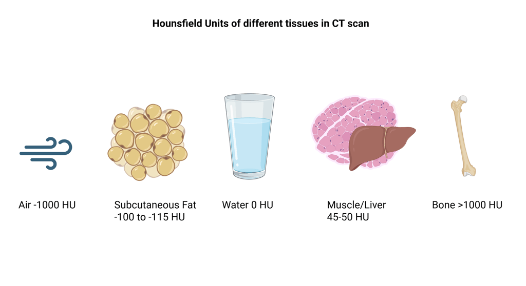
Non-contrast (unenhanced) CT remains the most reliable first-line imaging method for characterizing adrenal tumors. Hounsfield units (HU) provide a standardized numerical measure of tissue density on CT, reflecting X-ray attenuation relative to water (0 HU), with air defined at −1000 HU.

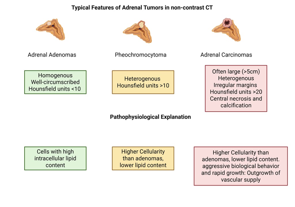
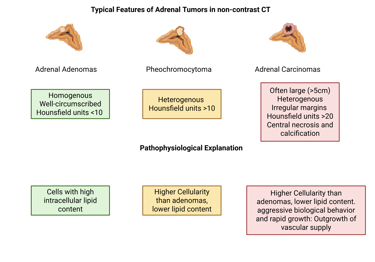
Adrenal lesions show characteristic density patterns on unenhanced scans:
Adrenal adenomas are typically small, homogeneous, and well-defined with HU values below 10, owing to their high intracellular lipid content.
Adrenocortical carcinomas often present as large (>5 cm), irregular, heterogeneous masses with HU values above 20, and frequently show central necrosis or hemorrhage due to rapid growth and limited vascular support.
Pheochromocytomas generally have HU >10 and appear heterogeneous, reflecting dense cellularity and low lipid content, which often places them in the indeterminate range on non-contrast imaging.
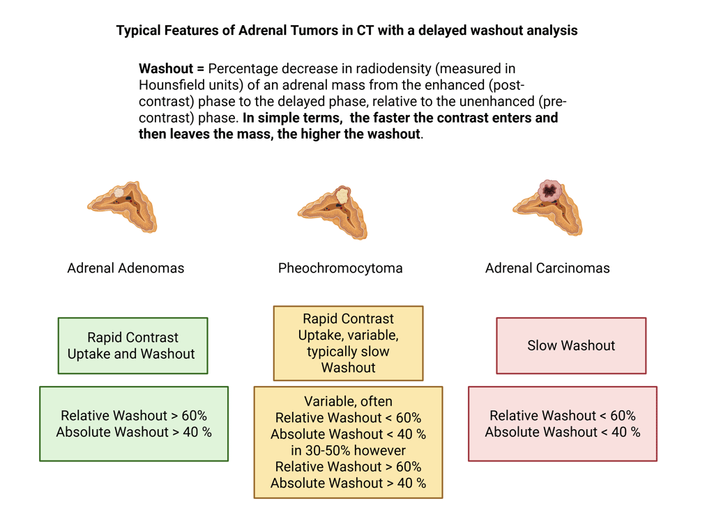
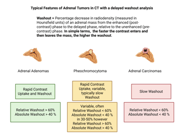
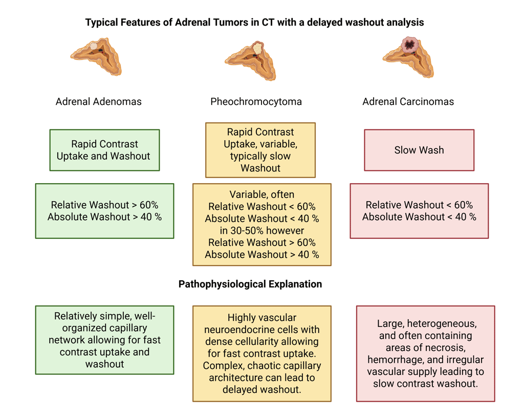
Further lesion characterization is achieved using multiphase CT, including non-contrast, portal venous (≈ 60–70 s post-contrast), and delayed (≈ 10–15 min post-contrast) phases. From these, washout is calculated using HU values across time points:
In clinical practice, washout CT serves as a second-line test when non-contrast findings are inconclusive. A relative washout >40% or absolute washout >60% strongly suggests a benign lipid-rich adenoma. In contrast, adrenocortical carcinomas and pheochromocytomas typically show slower or variable washout (<40% relative, <60% absolute), although up to 30–50% of pheochromocytomas can demonstrate rapid washout similar to adenomas.
The physiologic basis of these imaging differences lies in tumor composition and vascular architecture.
Adenomas contain abundant intracellular lipid and possess a simple, well-organized capillary network, leading to rapid contrast uptake and clearance (high washout).
Pheochromocytomas consist of densely cellular, highly vascular neuroendocrine tissue. Although they enhance strongly, their complex microvasculature and extracellular matrix slow contrast clearance.
Adrenocortical carcinomas, being heterogeneous and necrotic with disorganized vasculature, exhibit delayed and incomplete washout.

Take Home Messages
CT imaging features of adrenal tumors reflect their underlying biology; tissue composition and vascular structure determine attenuation and contrast behavior.
Lipid-rich adenomas appear homogeneous with low attenuation (<10 HU) due to abundant intracellular fat.
Adrenocortical carcinomas are large, heterogeneous, and irregular, showing high HU values, necrosis, and slow contrast washout because of poor vascular organization.
Pheochromocytomas are highly vascular and cellular, leading to high attenuation and variable washout, often mimicking malignant lesions.
Washout analysis helps differentiate benign from malignant lesions; rapid washout suggests adenoma, while slow or variable washout indicates other tumor types.
In summary: The CT appearance of an adrenal mass is not arbitrary, it mirrors its pathophysiologic makeup, offering key diagnostic clues.
References
All Illustrations were Created in https://BioRender.com
Rowe NE, Kumar R, Schieda N, Siddiqi F, McGregor T, McAlpine K, Violette P, Bathini V, Eng M, Izard J. Diagnosis, Management, and Follow-Up of the Incidentally Discovered Adrenal Mass: CUA Guideline Endorsed by the AUA. J Urol. 2023 Oct;210(4):590-599. doi: 10.1097/JU.0000000000003644. Epub 2023 Aug 9. PMID: 37556768.
Stahl KA, Sada A, Baytar Y, Dasyam AK, Maranchie JK, Fazeli PK, Seethala RS, McCoy KL, Yip L, Ramonell KM. Adrenal imaging features and associated pathologic diagnoses: A contemporary, longitudinal analysis. Surgery. 2025 Oct 9:109693. doi: 10.1016/j.surg.2025.109693. Epub ahead of print. PMID: 41033887.
Kebebew E. Adrenal Incidentaloma. N Engl J Med. 2021 Apr 22;384(16):1542-1551. doi: 10.1056/NEJMcp2031112. PMID: 33882207.
Ng L, Libertino JM. Adrenocortical carcinoma: diagnosis, evaluation and treatment. J Urol. 2003 Jan;169(1):5-11. doi: 10.1016/S0022-5347(05)64023-2. PMID: 12478091.
Schloetelburg W, Ebert I, Petritsch B, Weng AM, Dischinger U, Kircher S, Buck AK, Bley TA, Deutschbein T, Fassnacht M. Adrenal wash-out CT: moderate diagnostic value in distinguishing benign from malignant adrenal masses. Eur J Endocrinol. 2021 Dec 10;186(2):183-193. doi: 10.1530/EJE-21-0650. PMID: 34813495; PMCID: PMC8679842.
Hounsfield unit | Radiology Reference Article | Radiopaedia.org
© 2025 EndoCases. All rights reserved.
This platform is intended for medical professionals, particularly endocrinology residents, and is provided for educational purposes only. It supports learning and clinical reasoning but is not a substitute for professional medical advice or patient care. The information is general in nature and should be applied with appropriate clinical judgment and in accordance with local guidelines.
Portions of the text on this website were edited with the assistance of Artificial Intelligence to improve grammar and phrasing, as English is not my first language. All medical content, ideas for illustrations, and overall structure are original and based on the author’s own expertise and the cited medical literature. No AI tools were used to generate or influence the educational content itself.
All of the content is independent of my employer.
Use of this site implies acceptance of our Terms of Use
Contact us via E-Mail: contact@endo-cases.com