Clinical Implications of Endocrine Anatomy
The endocrine system comprises hormone-secreting cells that are either organized into anatomically distinct glands or embedded within tissues that serve additional physiological functions. The neuroendocrine system is composed of specialized cells that integrate neural and endocrine functions, characterized by their capacity to secrete hormones in response to neuronal stimulation. These cells are found both within classical endocrine organs, such as the adrenal medulla, and in organs primarily associated with non-endocrine functions. A prominent example of the latter is the gastrointestinal tract, which contains dispersed neuroendocrine cells collectively referred to as the enteroendocrine system. As a result, endocrine structures are widely distributed throughout the body and maintain close anatomical relationships with a variety of neighboring organs and systems.
These spatial relationships have significant clinical implications. Endocrine neoplasms, for instance, may manifest with symptoms not only attributable to hormonal dysregulation but also due to local mass effects on adjacent structures. A paradigmatic example is the pituitary adenoma, which, when enlarged, can exert pressure on the optic chiasm, leading to characteristic visual field defects such as bitemporal hemianopia. Conversely, pathological processes arising from neighboring non-endocrine structures can impair endocrine function. Sellar meningiomas, for example, may compress the pituitary gland, resulting in hypopituitarism. Moreover, diseases affecting organs that harbor endocrine or neuroendocrine cells can secondarily disrupt their hormonal output; for instance, pancreatic endocrine insufficiency leading to pancreoprivic diabetes mellitus in chronic pancreatitis, or pituitary dysfunction following traumatic brain injury.
A detailed understanding of the anatomical context of endocrine organs is therefore critical in both the diagnosis and management of endocrine disorders.

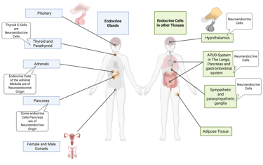
Illustration of Endocrine Organs and Cells of the human body. Created in https://BioRender.com
In endocrine glands comprising multiple hormone-producing cell lineages, these distinct cell types are often spatially organized into defined anatomical regions within the gland. For instance, in the thyroid gland, parafollicular C-cells, which secrete calcitonin, are located in the interfollicular spaces, distinct from the follicular cells responsible for thyroid hormone production. Similarly, in the anterior pituitary, hormone-producing cells such as somatotrophs, corticotrophs, and thyrotrophs are distributed in topographically distinct regions according to their developmental lineage and secretory function. Such spatial organization reflects the tightly regulated microenvironments necessary for endocrine signaling and feedback integration.
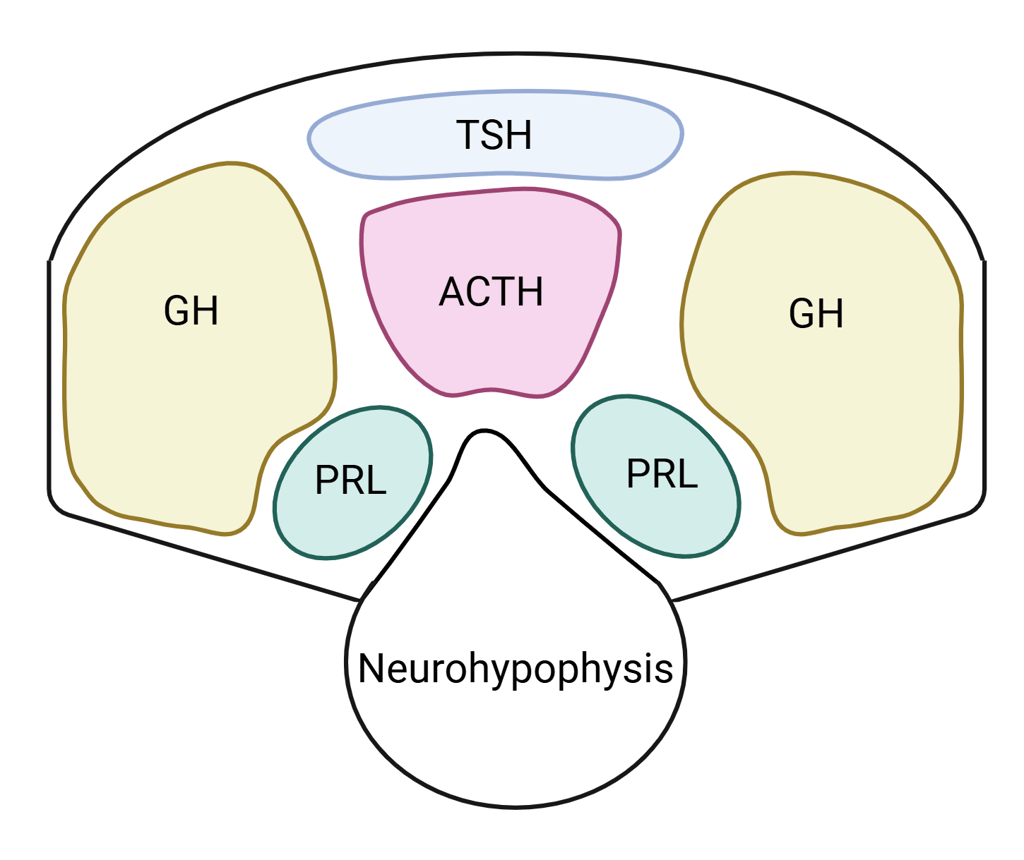

Illustration: Transverse section of the pituitary gland depicting the approximate locations of hormone-producing cell types—thyrotrophs (TSH), lactotrophs (PRL), corticotrophs (ACTH), and somatotrophs (GH). Note: Size proportions are illustrative and not exact.
Created in https://BioRender.com on the basis of: Cullingford DJ, Siafarikas A, Choong CS. Genetic Etiology of Congenital Hypopituitarism. [Updated 2023 Feb 5]. In: Feingold KR, Ahmed SF, Anawalt B, et al., editors. Endotext [Internet]. South Dartmouth (MA): MDText.com, Inc.; 2000-. Figure 7. [Cross-sectional representation of pituitary cell...]. Available from: https://www.ncbi.nlm.nih.gov/books/NBK586145/figure/neuroendo-congn-hypo.F7/
References
All Illustrations were created in https://BioRender.com
For References, visit the Section "References" in General Principles of Clinical Endocrinology
© 2025 EndoCases. All rights reserved.
This platform is intended for medical professionals, particularly endocrinology residents, and is provided for educational purposes only. It supports learning and clinical reasoning but is not a substitute for professional medical advice or patient care. The information is general in nature and should be applied with appropriate clinical judgment and in accordance with local guidelines.
Portions of the text on this website were edited with the assistance of Artificial Intelligence to improve grammar and phrasing, as English is not my first language. All medical content, ideas for illustrations, and overall structure are original and based on the author’s own expertise and the cited medical literature. No AI tools were used to generate or influence the educational content itself.
All of the content is independent of my employer.
Use of this site implies acceptance of our Terms of Use
Contact us via E-Mail: contact@endo-cases.com